+91 9939829148
 visionlasercentre@gmail.com
 +91 7761882875

Surgical Procedure
Tear Sac
All Treatments
Tear Sac
Understanding and Addressing Tear Duct Blockage
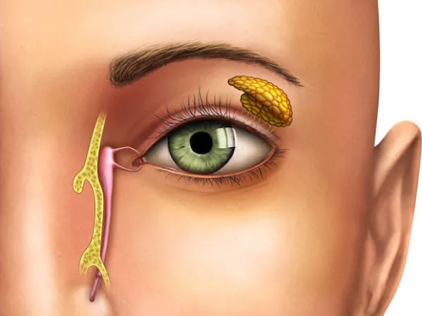
The tear sac, or lacrimal sac, is part of the tear drainage system located near the inner corner of the eye. It collects tears produced by the lacrimal glands and drains them into the nasal cavity through the tear ducts. When this drainage system becomes blocked or infected, it can lead to discomfort, excessive tearing, or serious infection. Our clinic specializes in diagnosing and treating issues related to the tear sac, ensuring that patients regain normal tear function and comfort.
What is a Tear Sac Blockage?
A blockage in the tear duct system, known as nasolacrimal duct obstruction (NLDO), prevents tears from draining properly, leading to a buildup in the tear sac. This condition can cause excessive tearing (epiphora), recurrent eye infections, and inflammation. Tear sac blockages are common in infants, but they can also affect adults, particularly after an injury or infection.
Symptoms of Tear Sac Blockage
- Excessive Tearing: A constant flow of tears that overflow onto the cheeks due to improper drainage.
- Recurrent Eye Infections: Frequent infections, including conjunctivitis, due to stagnant tears in the eye.
- Swelling and Redness: The area near the inner corner of the eye may appear swollen, red, and tender, especially if the tear sac becomes infected (dacryocystitis).
- Discharge from the Eye: Mucus or pus may discharge from the eye when pressure is applied to the tear sac.
- Blurred Vision: The buildup of tears can sometimes cause temporary blurring of vision.
Causes of Tear Sac Blockage
- Congenital Blockages: Many infants are born with a tear duct that hasn’t fully opened, leading to excessive tearing, which often resolves on its own within the first year of life.
- Age-Related Changes: As we age, the tear ducts can narrow, increasing the risk of blockages in adults.
- Infections or Inflammation: Infections in the nasal passages or sinuses can lead to inflammation and narrowing of the tear ducts.
- Trauma or Injury: Injuries to the face, particularly around the nose or eyes, can damage the tear drainage system.
- Tumors or Growths: In rare cases, growths or tumors in the nasal cavity or near the tear sac can block the tear ducts.
Diagnosis and Evaluation
A comprehensive eye exam and specific tests are necessary to diagnose a tear sac blockage. At our clinic, we conduct:
- Fluorescein Dye Test: A dye is placed in the eye to track how quickly tears drain. If the dye remains in the eye, it may indicate a blockage.
- Probing and Irrigation: A fine instrument is used to probe the tear duct, followed by irrigation to see if the blockage can be cleared.
- Dacryocystography: This imaging test uses a contrast dye injected into the tear sac, allowing detailed X-rays of the tear drainage system.
Treatment Options for Tear Sac Blockage
Treatment depends on the cause, age, and severity of the blockage. Common options include:
- Massage and Observation (for Infants): In many cases, a gentle massage of the tear sac can help open a partially blocked duct in infants. This non-invasive technique is combined with careful monitoring as the duct may open naturally over time.
- Antibiotics: If an infection is present, antibiotic eye drops or oral medications are prescribed to clear the infection before any further treatment is pursued.
- Probing (for Children): If a tear duct does not open naturally in infants, a simple procedure called tear duct probing can be performed. Under light sedation, a small probe is inserted into the duct to open the blockage.
- Dacryocystorhinostomy (DCR Surgery): For adults or chronic cases where the blockage persists, DCR surgery may be required. This procedure creates a new tear drainage pathway between the tear sac and the nasal cavity, bypassing the blockage. The surgery can be done externally or endoscopically, through the nose, with minimal scarring.
- Balloon Catheter Dilation: In this procedure, a small balloon is inserted into the blocked duct and inflated to widen the passage, allowing tears to drain normally. This method is less invasive than traditional surgery and can be used for both children and adults.
- Stenting or Intubation: A tiny tube or stent may be placed in the tear duct to keep the passage open while it heals. This is often done during surgery for more severe blockages.
Recovery and Follow-Up Care:
Recovery from tear sac treatments is generally quick, especially in infants. After surgery or procedures, most patients can return to normal activities within a few days. For surgical cases, there may be some mild bruising or swelling around the eye, but this typically resolves within a week. Post-treatment care includes:
- Antibiotic Eye Drops: To prevent infection and promote healing.
- Regular Follow-Ups: To ensure the tear duct remains open and the symptoms have resolved.
Frequently Asked Questions:
- Can tear sac blockages resolve on their own? In infants, many tear duct blockages resolve naturally within the first year of life. In adults, however, blockages typically require medical intervention.
- Is tear duct surgery painful? DCR surgery is typically performed under local or general anesthesia, so patients do not feel pain during the procedure. Any post-surgery discomfort is usually mild and manageable with over-the-counter pain relief.
- What happens if I don’t treat a blocked tear sac? If left untreated, a blocked tear sac can lead to chronic infections, swelling, and pain. In severe cases, untreated blockages can result in a serious infection that spreads to nearby tissues.
Schedule Your Tear Sac Consultation
If you are experiencing symptoms of a tear sac blockage, such as excessive tearing or frequent eye infections, it’s important to seek early treatment. Contact us today to schedule a consultation with our specialists. We provide personalized care to restore normal tear drainage and alleviate discomfort.